Percutaneous Needle Tenotomy (PNT)
What Is Ultrasound Guided Percutaneous Needle Tenotomy (PNT)?
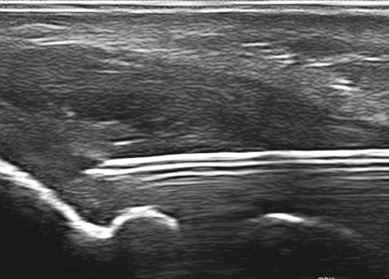
It is a minimally invasive intervention performed under direct ultrasound guidance, utilized in the treatment of chronic tendon problems known as tendinosis or tendinopathy. A small needle is introduced through the skin, and small multiple needling are performed in the damaged tendon, ultimately creating an enhanced healing response resulting in tendon repair. It is a cost effective intervention with excellent results, minimal morbidity and early return to function.
What Is Tendinosis/Tendinopathy?
It is a chronic degenerative condition caused by excessive strain, overuse and micro trauma which results in an abnormal tendon, not able to heal itself. There are other associated conditions which may predispose patients to developed abnormal tendon changes such as: calcification, anatomic mal-alignment, inadequate mechanics, poor training, inadequate rest or medication related (certain antibiotics).
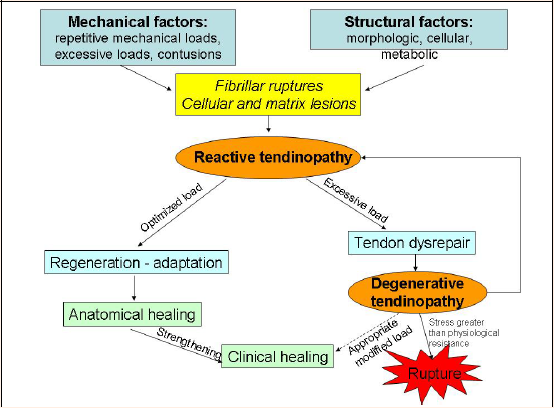
Cook JL, Purdam CR. Is tendon pathology a continuum? A pathology model to explain the clinical presentation of load-induced tendinopathy. BJSM 2009;43:409-416
When Is the Procedure Indicated?
Most of the time, tendon related problems can be treated conservatively. Usually physical therapy, orthotics/splinting, proper training methods, adequate rest, analgesics and rehabilitation, can improve or resolve the symptoms altogether. When conservative therapy fails and there is significant morbidity, PNT should be considered.
Which Are the Most Common Treated Conditions?
Tennis elbow, Golfer’s elbow, plantar fasciitis, gluteus tendinopathy, hamstring tendinopathy, Achilles tendinopathy, rotator cuff tendinosis and others.
How Is the Procedure Performed?
Percutaneous needle tenotomy is usually an office based intervention, performed under local anesthesia. The skin is cleansed and under ultrasound guidance the affected tendon site is identified and marked. Local anesthetic is infiltrated in the skin and over the affected tendon. A small needle is then advanced through the tendon into the bony origin. Multiple perforations area carried under ultrasound guidance. The needle is removed and a bandage applied. The procedure usually takes 20 minutes.
Post Procedure Instructions
Active range of motion is encouraged during the first 48 hours. Mild analgesics are recommended along with cold compresses. The use of non-steroidal should be avoided for 2 weeks post procedure. Avoidance of forceful resistance exercises is recommended. Physical therapy is strongly recommended post-procedure. A gradual tendon eccentric loading program should be started 1 week post procedure.
References: